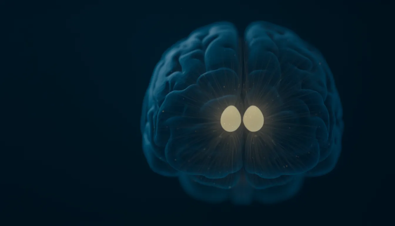
Thalamus
Also known as: thalamus function, thalamus location, what does the thalamus do, thalamic nuclei, thalamocortical, pulvinar, lateral geniculate nucleus, thalamic reticular nucleus, mediodorsal thalamus
In one sentenceThe thalamus is a pair of egg-shaped masses of grey matter at the center of the brain that relays sensory and motor signals to the cortex and helps regulate sleep, alertness and attention.
What does the thalamus do?
The thalamus is a pair of egg-shaped masses of grey matter at the center of the brain, best known as the brain’s relay station. Signals from all the senses except smell, along with signals involved in movement, pass through it before they reach the cerebral cortex, the brain’s outer layer, where they are processed further. The thalamus also takes part in sleep and wakefulness, attention, learning and memory.
The thalamus has long been described mainly as a relay that passes information on to the cortex. Research has since shown that it also controls whether and in what form that information gets through, links areas of the cortex to one another, and helps sustain activity in thinking areas of the cortex; a 2017 study concluded that the thalamus has a much broader role in cognition than previously thought. The sections below set out where it is, how it is built, and the research behind each of these roles.
Location and name
The thalamus lies above the brainstem in the middle of the brain, between the brainstem and the cerebral cortex. There are two thalami side by side, one in each half of the brain; Cleveland Clinic describes them as looking like a pair of eggs, each about 3 to 4 centimeters (1 to 1.5 inches) long. The Queensland Brain Institute describes the thalamus as two lobes of grey matter tucked away under the cerebral cortex. The hypothalamus lies right underneath it.
Both structures develop from the same part of the embryonic brain. By the fifth week of human development the front-most of the brain’s three early vesicles has divided into the telencephalon, which becomes the cerebral hemispheres, and the diencephalon, which goes on to form the thalamus and the hypothalamus. The development of the thalamus comes before that of the telencephalon.
The thalamus was first described by the physician and philosopher Galen in the second century AD. Its name comes from a Greek word meaning “inner chamber” or “anteroom”.
Nuclei
The thalamus is not one uniform structure but a collection of nuclei, clusters of neurons, each with its own inputs and outputs, most of which send information to the cerebral cortex. Counts vary with the atlas used: a 2025 review divides it into around 30 nuclei, while the StatPearls entry on thalamic nuclei gives approximately 60. The divisions are not easy to see on MRI scans. Some of the best known are:
- Ventral posterolateral and ventral posteromedial nuclei: relay sensory information from the limbs and trunk and from the head and face, respectively, to the somatosensory cortex.
- Lateral geniculate and medial geniculate nuclei: receive direct input from the visual and auditory systems and project to the visual and auditory cortex.
- Ventral anterior and ventral lateral nuclei: the parts most connected to the motor system, receiving input from the cerebellum.
- Pulvinar: the largest thalamic nucleus, with extensive connections to the posterior parietal, occipital, insular and cingulate cortex. It is thought to be involved in combining information from several senses and in visual attention, and of all thalamic nuclei it has grown the most in size over evolution.
- Dorsomedial nucleus: the largest nucleus on the medial (inner) surface, with extensive projections to the prefrontal cortex. It receives input from the limbic system, including the amygdala, and is involved in emotion, memory and level of consciousness.
- Anterior nucleus: part of a memory-related circuit with the hippocampus, described below.
- Reticular nucleus: a layer that covers the outer side of the thalamus like a blanket. It is the only thalamic nucleus that sends no excitatory connections outside the thalamus; its neurons project only back onto the thalamus, and their connections are strictly inhibitory.
- Intralaminar nuclei: smaller nuclei encircled by the internal medullary lamina, a layer that also separates some of the larger nuclei. One of them, the centromedian nucleus, projects mainly to the striatum, part of the basal ganglia, and is part of the reticular activating system, which regulates wakefulness, arousal and consciousness.
Two kinds of neurons
The thalamus contains two basic types of neurons. Projection neurons are excitatory, use the neurotransmitter glutamate, and send their axons out of the thalamus to the cortex, the striatum and the amygdala; on the way out they send side branches to the reticular nucleus. Unlike neurons in the cortex, which receive abundant excitatory input from their neighbors, thalamic projection neurons receive no excitatory connections from other thalamic neurons. The second type, inhibitory interneurons that use GABA, stay within their own nucleus and are densely connected with the projection neurons and with each other.
More than a relay: drivers and modulators
In 1998 S. Murray Sherman and R. W. Guillery proposed that the inputs reaching a thalamic neuron fall into two classes. “Drivers” carry the information itself, transmitting a neuron’s receptive field properties, that is, the stimuli it responds to; the input from the retina to the lateral geniculate nucleus is an example. “Modulators” instead alter the probability of certain aspects of that transmission. They suggested that any one cell is likely to have few drivers but many modulators, and that drivers act quickly through ionotropic receptors while modulators can also act through metabotropic receptors with slower, longer-lasting effects.
According to a 2025 review, modulatory inputs to the thalamus come from layer 6 of the cortex, the basal ganglia, the inhibitory neurons of the thalamus itself, the cerebellum and the reticular formation of the brainstem, and use transmitters including glutamate, GABA, serotonin, dopamine, acetylcholine and norepinephrine. They far outnumber the drivers and determine whether, and in what form, the content of the driver input reaches the cortex.
In a 2016 review Sherman distinguished two kinds of thalamic relays by where their drivers come from. First-order relays, such as the lateral geniculate nucleus, receive their driver input from below the cortex, for example from the retina. Higher-order relays, such as the pulvinar, receive their driver input from layer 5 of the cortex itself, and so form cortex–thalamus–cortex circuits. Sherman argued that direct connections between cortical areas are often paralleled by such routes through the thalamus, an aspect of how the cortex works that had been little appreciated.
Sleep, wakefulness and firing modes
Thalamic projection neurons have two firing modes. In tonic mode, when the cell is relatively depolarized, it fires a train of action potentials whose rate reflects the strength of its input, so information is passed on faithfully. Burst mode depends on T-type calcium channels: when the cell has been strongly hyperpolarized, a small depolarization opens these channels, and the inflow of calcium triggers a short burst of action potentials that does not follow the input faithfully. Burst mode was originally described in drowsiness and sleep, when thalamic neurons are mostly in this mode, which effectively blocks the transmission of specific sensory input. Burst mode does still carry some information and can act as a sensitive detector of change.
In a 1993 review in Science, Mircea Steriade, David McCormick and Terrence Sejnowski described sleep as marked by synchronized activity among billions of connected neurons in thalamocortical systems. On waking, a series of neuromodulatory transmitter systems become active, blocking the slow oscillations of sleep, bringing in fast rhythms and allowing the brain to recover full responsiveness. Sleep spindles, a type of sleep rhythm, are thought to arise from a loop between the reticular nucleus and the other thalamic nuclei. The Queensland Brain Institute lists sleep control among the thalamus’s roles, and Cleveland Clinic describes it as playing a role in keeping a person awake and alert.
Attention
In 1984 Francis Crick proposed what he called the searchlight hypothesis. Treisman and others had proposed that attention works like an internal searchlight; Crick suggested that this searchlight is controlled by the reticular complex of the thalamus, and that it is expressed as rapid bursts of firing in a subset of thalamic neurons.
Later experiments tested the thalamus’s part in attention more directly:
- Pulvinar in monkeys (2012). Yuri Saalmann, Sabine Kastner and colleagues mapped the connections between the pulvinar and visual areas of the cortex with diffusion tensor imaging, then recorded from these connected sites at the same time while monkeys performed a visual attention task. The pulvinar synchronized activity between connected cortical areas according to where the animals were paying attention, which the authors took as evidence that the thalamus helps regulate how information is passed between areas of the visual cortex.
- Reticular nucleus in mice (2015). Ralf Wimmer, Michael Halassa and colleagues trained mice to choose between conflicting visual and auditory cues. Briefly disrupting the prefrontal cortex optogenetically impaired the choice, and the behavior depended on the prefrontal cortex’s interactions with the sensory thalamus rather than the sensory cortex. Neurons in the visual part of the reticular nucleus changed their firing, depending on the prefrontal cortex, in a way that predicted which sense the mouse would select, and they controlled the gain of the visual thalamus through inhibition. The authors proposed that the prefrontal cortex selects sensory input by biasing reticular subnetworks.
Thinking and working memory
A group of studies has examined the two-way connections between the mediodorsal thalamus and the prefrontal cortex in mice.
In 2017 L. Ian Schmitt, Michael Halassa and colleagues studied how the mouse prefrontal cortex represents two rules used to guide attention. The mediodorsal thalamus sustained these rule representations without itself carrying the rule information: its input amplified connections within the prefrontal cortex, allowing rule-specific sequences of neural activity to emerge. Broadly increasing prefrontal excitability made the representations less specific and performance worse, while increasing mediodorsal excitability improved both. The authors described this as thalamic control of functional connectivity in the cortex.
Also in 2017, Scott Bolkan, Christoph Kellendonk and colleagues blocked specific pathways between the two regions during a working memory task. The pathway from the mediodorsal thalamus to the prefrontal cortex supported holding information in mind during a delay, while the reverse pathway supported the choice that followed. Prefrontal neurons that fired more during the delay needed thalamic input to keep doing so, and increasing the excitability of the mediodorsal thalamus improved task performance. The authors noted that reduced connectivity between these two regions has been observed in people with schizophrenia, who also show impairments in working memory.
A 2017 review by Halassa and Kastner summarized such findings from rodents and primates: in addition to relaying information to or between cortical areas, thalamic circuits shift and sustain the interactions within and across cortical areas, rapidly coordinating separate cortical computations into networks suited to the task at hand.
Memory
The anterior nucleus is part of the limbic circuit historically known as the Papez circuit. The pathway runs from the hippocampus through the fornix to the mammillary bodies, which project to the anterior nucleus through the mammillothalamic tract. The anterior nucleus in turn connects to the anterior cingulate cortex, which projects to the entorhinal cortex and back to the hippocampus. The anterior nucleus has also been implicated in organizing sleep spindles and in coordinating sleep rhythms during memory consolidation.
In a 1999 paper John Aggleton and Malcolm Brown argued that the usual distinction between amnesia caused by damage to the temporal lobe and amnesia caused by damage to the diencephalon is of limited value. A common feature of anterograde amnesia, the inability to form new memories, is damage to part of an “extended hippocampal system” made up of the hippocampus, the fornix, the mammillary bodies and the anterior thalamic nuclei, which they proposed is needed for encoding and later recalling episodic memories. They traced this view back to Delay and Brion in 1969. Judging whether something is merely familiar, they proposed, depends on a separate system involving the perirhinal cortex and the medial dorsal nucleus of the thalamus.
When the thalamus is damaged
Because so many pathways pass through the thalamus, damage to it can affect many functions. The main causes include stroke and tumors. Depending on exactly where it is, damage to the blood supply of the thalamus can cause memory problems, reduced consciousness or coma, aphasia (difficulty with language), loss of coordination on one side of the body, a pain syndrome and loss of sensation. Damage to the dorsomedial nucleus on both sides can be a cause of coma.
Several rare conditions involve the thalamus. In Korsakoff syndrome, which is linked to vitamin B1 deficiency due to alcohol use disorder, the brain shows widespread atrophy with the most marked degeneration in the anterior thalamic nucleus. Fatal familial insomnia, a rare inherited prion disease, is thought to destroy the dorsomedial and anterior nuclei preferentially; insomnia, with a lack of slow-wave sleep, is one of its earliest signs.
Related entries
- Hypothalamus — The hypothalamus is a small region at the base of the brain, below the thalamus, that keeps the body's internal state stable through the autonomic nervous system and hormones.
- Prefrontal cortex — The prefrontal cortex, just behind the forehead, runs executive functions such as planning, impulse control and working memory, and matures last.
- Working memory — Working memory holds information in mind for seconds while you manipulate it, supporting mental arithmetic, conversation and planning.
- Hippocampus — The hippocampus is a structure deep in the temporal lobe that is central to turning new events and facts into long-term memories and to remembering places.
References
- Forebrain. Queensland Brain Institute, The University of Queensland
- Thalamus. Cleveland Clinic, medically reviewed, last updated 5 February 2026
- The thalamus: Structure, function, and neurotherapeutics. Marcuse LV, Langan M, Hof PR et al. Neurotherapeutics 22, e00550 (2025)
- Neuroanatomy, Thalamic Nuclei. Sheridan N, Tadi P. StatPearls, NCBI Bookshelf
- On the actions that one nerve cell can have on another: distinguishing "drivers" from "modulators". Sherman SM, Guillery RW. PNAS 95, 7121–7126 (1998)
- Thalamus plays a central role in ongoing cortical functioning. Sherman SM. Nature Neuroscience 19, 533–541 (2016)
- Thalamocortical oscillations in the sleeping and aroused brain. Steriade M, McCormick DA, Sejnowski TJ. Science 262, 679–685 (1993)
- Function of the thalamic reticular complex: the searchlight hypothesis. Crick F. PNAS 81, 4586–4590 (1984)
- The pulvinar regulates information transmission between cortical areas based on attention demands. Saalmann YB, Pinsk MA, Wang L, Li X, Kastner S. Science 337, 753–756 (2012)
- Thalamic control of sensory selection in divided attention. Wimmer RD, Schmitt LI, Davidson TJ, Nakajima M, Deisseroth K, Halassa MM. Nature 526, 705–709 (2015)
- Thalamic functions in distributed cognitive control. Halassa MM, Kastner S. Nature Neuroscience 20, 1669–1679 (2017)
- Thalamic amplification of cortical connectivity sustains attentional control. Schmitt LI, Wimmer RD, Nakajima M, Happ M, Mofakham S, Halassa MM. Nature 545, 219–223 (2017)
- Thalamic projections sustain prefrontal activity during working memory maintenance. Bolkan SS, Stujenske JM, Parnaudeau S et al. Nature Neuroscience 20, 987–996 (2017)
- Episodic memory, amnesia, and the hippocampal–anterior thalamic axis. Aggleton JP, Brown MW. Behavioral and Brain Sciences 22, 425–444 (1999)