Cerebellum
Also known as: little brain, cerebellar cortex, cerebellum function, cerebellum location
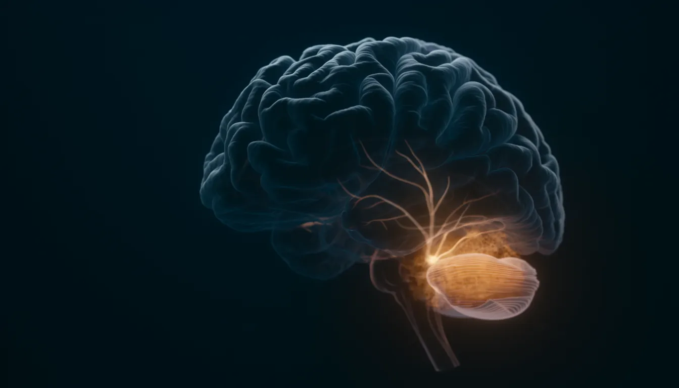
In one sentenceThe cerebellum, or "little brain", sits at the back of the head behind the brainstem. It coordinates movement and balance, learns motor skills and also links to thinking areas of the cortex.
What does the cerebellum do?
The cerebellum coordinates sensation with the responses of the muscles, enabling most voluntary movements, and it uses signals from the inner ear to help maintain balance and posture. It also learns: it adjusts movements that miss their target, and it predicts what our own movements will feel like. Its connections reach beyond the motor system, to parts of the prefrontal cortex involved in working memory. The evidence for each point is set out below.
Location and structure
The cerebellum, Latin for “little brain”, sits at the lower back of the brain, behind the pons and the rest of the brainstem, and is part of the hindbrain. Like the cerebral cortex, it has two hemispheres, with a dense layer of grey matter around an inner region of white matter. Its best-known cells are the Purkinje cells, whose highly complex dendritic branches let them process many signals at once.
Small as it looks, the cerebellum holds most of the brain’s neurons. Counts of cells in adult human brains, summarized by Suzana Herculano-Houzel in 2009, found about 86 billion neurons in total, about 69 billion of them in a cerebellum weighing 154 g, roughly a tenth of the brain’s mass. The cerebral cortex, by contrast, makes up 82% of brain mass but holds only 19% of its neurons. Most cerebellar neurons are tiny granule cells; a 2017 study put their number at about 60 billion, more than all other neurons in the brain combined.
The cerebellar surface is also much more tightly folded than the cerebral cortex. In 2020 Martin Sereno and colleagues reconstructed it down to its individual small folds (folia) from high-resolution MRI scans of post-mortem brains. Unfolded, its surface measured about 1,590 cm², equal to 78% of the surface area of the human neocortex, and formed a strip about 10 cm wide and almost 1 m long. In the macaque monkey the same comparison gave about 33%.
The basic circuit
Two kinds of input fibres reach the cerebellar cortex. Mossy fibres carry information about the context of a movement to the granule cells, which pass it on to Purkinje cells through parallel fibres that cross the Purkinje cells’ sheet-like dendritic trees at right angles. Each Purkinje cell also receives a second, very different input: a climbing fibre from a structure called the inferior olive. The cerebellum’s projections to the cerebral cortex start in a set of cerebellar nuclei, including the dentate nuclei, its major output nuclei, and pass through the thalamus.
Learning movements
In 1969 David Marr proposed a theory in which the cerebellum learns motor skills. In his model, a climbing fibre signals an instruction for an elementary movement. When it fires together with parallel fibres, the parallel-fibre synapses onto that Purkinje cell are strengthened, so that after practice the context alone is enough to fire the Purkinje cell. James Albus published a related theory in 1971, and the shared idea became known as the Marr–Albus model.
In 1982 Masao Ito and Masanobu Kano stimulated parallel fibres and climbing fibres together in the cerebellar cortex of rabbits and found a depression of parallel fibre to Purkinje cell transmission that lasted at least an hour; stimulating either input alone had no such effect. The change ran in the opposite direction to Marr’s predicted strengthening, but the authors concluded that it supported the plasticity assumption of the Marr–Albus model: that these synapses can be modified.
Other experiments tied specific kinds of learning to the cerebellum:
- Conditioned eyeblinks. In 1984 David McCormick and Richard Thompson trained rabbits to blink in response to a cue. Lesions of two cerebellar nuclei, the dentate and interpositus, on the same side abolished the learned blink, while lesions of the cerebellar cortex did not. Neurons in those nuclei responded in ways related to the learning, and stimulating them produced the blink.
- Adapting to prism glasses. In 1996 Martin, Thach and colleagues asked people to throw balls of clay at a target while wearing wedge prism glasses that bent their line of sight. Healthy volunteers first threw in the direction the prisms bent their gaze but corrected their aim over repeated throws. Patients with damage to certain parts of the olivocerebellar system adapted poorly or not at all. Some patients had little or no loss of coordination but could not adapt, while others had the reverse pattern, suggesting that the two depend on partly different parts of the system.
Predicting the results of our own movements
A leading idea is that the cerebellum contains internal models of the body. In a 1998 review, Daniel Wolpert, Chris Miall and Mitsuo Kawato described two kinds: inverse models, which provide the commands needed to produce a desired movement, and forward models, which predict the consequences of a movement and can be used to overcome the time delays of control based on sensory feedback.
Prediction may also explain why a touch you produce yourself feels less ticklish than the same touch from outside. In 1998 Blakemore, Wolpert and Chris Frith used fMRI to compare the two. The somatosensory cortex responded more to the externally produced touch. In the cerebellum, a movement that produced a touch was associated with less activity than a movement that did not. The authors interpreted this as the cerebellum predicting the sensory result of a movement and supplying a signal used to cancel the response to self-produced touch.
Beyond movement
Whether the cerebellum takes part in more than movement was long a controversial question. Several lines of evidence now link it to other functions.
- Anatomy. In 1994 Frank Middleton and Peter Strick traced connections in primates with a virus that travels from neuron to neuron. Neurons in the dentate nucleus projected through the thalamus to area 46 of the prefrontal cortex in the frontal lobe, a region involved in spatial working memory; the same study found output from the basal ganglia reaching this area. A 2009 review by Strick and colleagues concluded that cerebellar output reaches prefrontal and posterior parietal areas as well as motor areas, organized as closed loops: the cortical area that receives a given output channel is also a major source of input to it. The review also listed the wide range of tasks in which imaging studies had found cerebellar activity, including attention, executive control, language, working memory, learning, pain and emotion.
- Patients. In 1998 Schmahmann and Sherman examined 20 patients whose disease was confined to the cerebellum. Those with lesions of the posterior lobe and the vermis, the cerebellum’s midline, showed changes in executive functions such as planning, abstract reasoning and working memory, in spatial cognition, in personality and in language. Lesions of the anterior lobe produced only minor changes of this kind. The authors named the pattern the cerebellar cognitive affective syndrome and suggested that it reflects disrupted cerebellar modulation of the circuits linking the cerebellum with prefrontal, posterior parietal, superior temporal and limbic areas of the cortex.
- Brain imaging. In 2011 Randy Buckner and colleagues used resting-state functional connectivity MRI in 1,000 people to estimate which part of the cerebral cortex each point of the cerebellum is principally linked to. Most of the human cerebellum mapped to association areas of the cortex, and the cerebellum contained at least two large maps of the whole cerebral cortex, one in its anterior and one in its posterior part, arranged as mirror images.
- Reward. In 2017 Mark Wagner, Liqun Luo and colleagues imaged granule cells in mice learning to earn a sugar-water reward. Some granule cells responded to the reward or to its omission, and others came to signal that a reward was expected. Granule cells had previously been known to encode only sensory and motor context.
Related entries
- Frontal lobe — The frontal lobe is the front part of each cerebral hemisphere. It controls voluntary movement, produces speech and, through the prefrontal cortex, plans and regulates behavior.
- Prefrontal cortex — The prefrontal cortex, just behind the forehead, runs executive functions such as planning, impulse control and working memory, and matures last.
- Basal ganglia — The basal ganglia are deep brain nuclei that loop with the cortex and thalamus to start and select movements and to learn habits.
References
- The hindbrain. Queensland Brain Institute, The University of Queensland
- The human brain in numbers: a linearly scaled-up primate brain. Herculano-Houzel S. Frontiers in Human Neuroscience 3, 31 (2009)
- The human cerebellum has almost 80% of the surface area of the neocortex. Sereno MI, Diedrichsen J, Tachrount M, Testa-Silva G, d'Arceuil H, De Zeeuw C. PNAS 117, 19538–19543 (2020)
- A theory of cerebellar cortex. Marr D. The Journal of Physiology 202, 437–470 (1969)
- A theory of cerebellar function. Albus JS. Mathematical Biosciences 10, 25–61 (1971)
- Long-lasting depression of parallel fiber-Purkinje cell transmission induced by conjunctive stimulation of parallel fibers and climbing fibers in the cerebellar cortex. Ito M, Kano M. Neuroscience Letters 33, 253–258 (1982)
- Cerebellum: essential involvement in the classically conditioned eyelid response. McCormick DA, Thompson RF. Science 223, 296–299 (1984)
- Throwing while looking through prisms. I. Focal olivocerebellar lesions impair adaptation. Martin TA, Keating JG, Goodkin HP, Bastian AJ, Thach WT. Brain 119, 1183–1198 (1996)
- Internal models in the cerebellum. Wolpert DM, Miall RC, Kawato M. Trends in Cognitive Sciences 2, 338–347 (1998)
- Central cancellation of self-produced tickle sensation. Blakemore SJ, Wolpert DM, Frith CD. Nature Neuroscience 1, 635–640 (1998)
- Anatomical evidence for cerebellar and basal ganglia involvement in higher cognitive function. Middleton FA, Strick PL. Science 266, 458–461 (1994)
- Cerebellum and nonmotor function. Strick PL, Dum RP, Fiez JA. Annual Review of Neuroscience 32, 413–434 (2009)
- The cerebellar cognitive affective syndrome. Schmahmann JD, Sherman JC. Brain 121, 561–579 (1998)
- The organization of the human cerebellum estimated by intrinsic functional connectivity. Buckner RL, Krienen FM, Castellanos A, Diaz JC, Yeo BT. Journal of Neurophysiology 106, 2322–2345 (2011)
- Cerebellar granule cells encode the expectation of reward. Wagner MJ, Kim TH, Savall J, Schnitzer MJ, Luo L. Nature 544, 96–100 (2017)