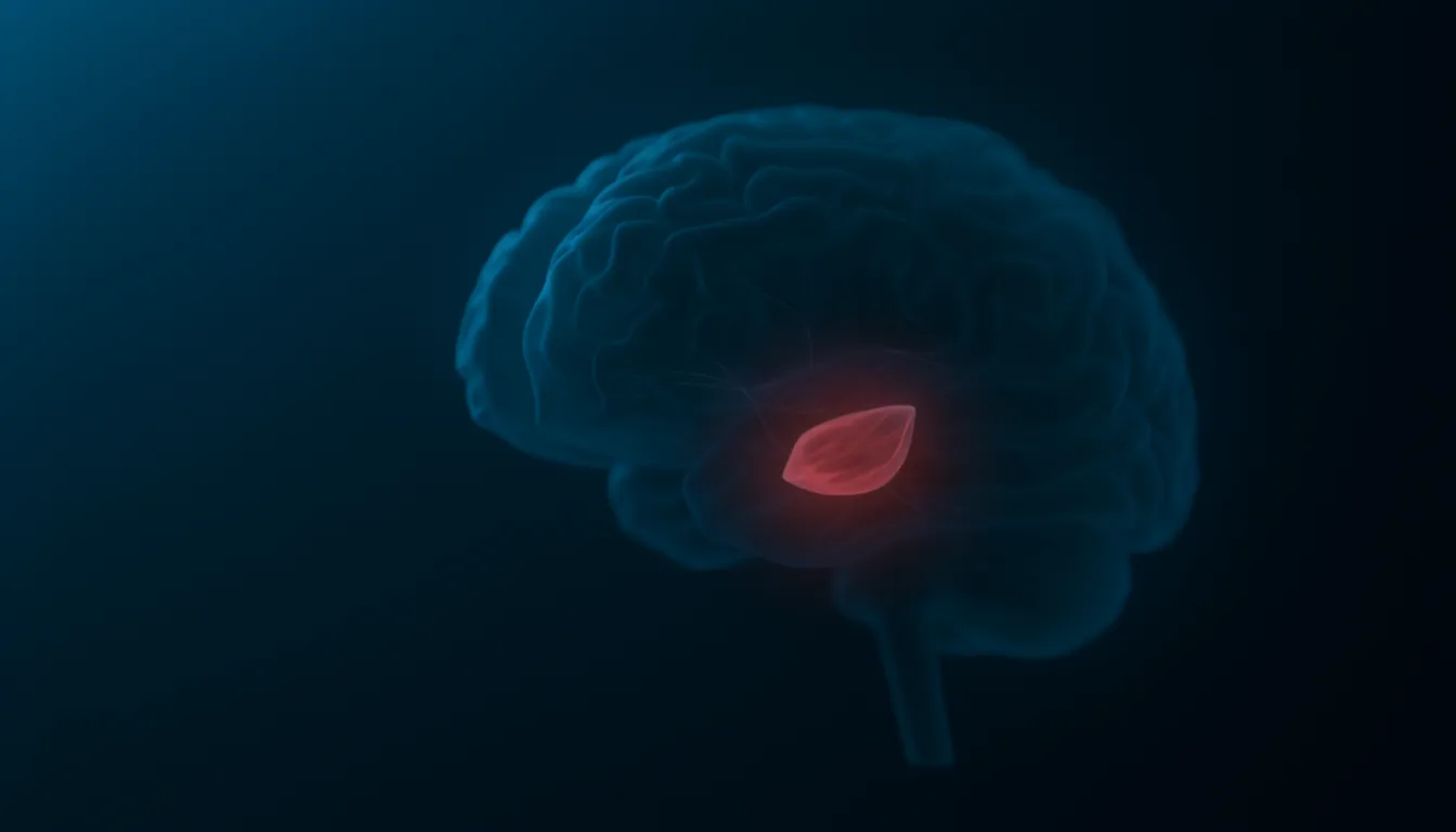
Amygdala
Also known as: amygdalae, amygdaloid complex, amygdala function, amygdala location
In one sentenceThe amygdala is an almond-shaped cluster of nuclei deep in each temporal lobe, next to the hippocampus. It links sights and sounds with emotional value, above all threat.
What does the amygdala do?
The amygdala links things we see and hear with their emotional value. It is best known for threat: it is needed to learn that a sound or image predicts danger, to recognize fear in other people’s faces, and to trigger fear in response to dangers in the outside world. It also helps emotionally charged events stick in memory and responds to rewards as well as threats. The evidence for each point is set out below.
Location and name
There are two amygdalae, a left and a right, one deep inside each temporal lobe right next to the hippocampus. The name refers to the structure’s almond-like shape.
Although small, the amygdala is not a single unit but a group of interconnected nuclei. Two parts are studied most. The basolateral complex receives information about the outside world from sensory areas of the thalamus and cortex and exchanges signals with the prefrontal cortex and the hippocampus. The central nucleus is one of the main routes by which amygdala signals are turned into behavior.
Early clues
The first hints came from monkeys. In 1888 Brown and Schäfer removed the temporal lobes of a rhesus monkey and reported reduced aggression, fear and defensive behavior, and Klüver and Bucy later reported similar changes. Those changes were mixed with effects now attributed to other brain regions. In 1956 Lawrence Weiskrantz removed the amygdala itself and found that the animals had difficulty learning responses to cues that predicted a shock.
Learning what to fear
Much of what is known about the amygdala comes from fear conditioning, in which a neutral cue is paired with an unpleasant event such as a mild shock until the cue alone produces signs of fear. Reviewing this work in 2000, Joseph LeDoux identified the amygdala as an important component of the system that acquires, stores and expresses fear memories.
A 1995 study by Antoine Bechara, Antonio Damasio and colleagues separated this kind of learning from conscious memory. A patient with damage to both amygdalae, S.M. (see below), did not acquire conditioned autonomic (bodily) responses to a cue but did learn which cue had been paired with the unpleasant stimulus. A patient with damage to both hippocampi showed the reverse: the conditioned response appeared, but the facts were not learned.
The patient S.M.
Much human evidence comes from a rare patient known as S.M., who has damage confined to both amygdalae caused by Urbach-Wiethe disease. S.M. is the most intensively studied case of its kind.
- In 1994 Ralph Adolphs and colleagues found that S.M. had difficulty recognizing fear in facial expressions but could still recognize who a face belonged to.
- In 2011 Justin Feinstein and colleagues tried to provoke fear in S.M. with live snakes and spiders, a haunted house and frightening films. S.M. never showed fear and never reported more than minimal fear, while still showing and reporting other basic emotions.
- In 2009 Daniel Kennedy and colleagues found that S.M. lacked any sense of personal space, and that in healthy people the amygdala became active when another person stood very close.
Not the whole story of fear
The amygdala is often called the brain’s fear center, but research points to a narrower role. In 2013 Feinstein and colleagues had three patients with damage to both amygdalae, including S.M., inhale air containing 35% carbon dioxide. All three felt fear and had panic attacks; the authors noted that, to their knowledge, it was the first time S.M. had felt fear in any setting since childhood. They concluded that the amygdala is not required for fear and panic, and distinguished fear triggered by external threats from fear triggered internally, as by carbon dioxide.
LeDoux has argued in a similar direction that the mechanisms that detect and respond to threats are not the same as those that give rise to the conscious feeling of fear. He proposed describing so-called fear conditioning in terms of circuits that operate without awareness and contribute only indirectly to conscious fear.
Emotional memories
The amygdala also strengthens memories. In 1996 Larry Cahill, James McGaugh and colleagues measured brain activity with PET while people watched emotionally arousing and neutral film clips. Three weeks later, activity in the right amygdala during the emotional clips was closely correlated with how many of those clips people recalled; there was no such link for the neutral clips. Reviewing animal and human studies in 2004, McGaugh concluded that the amygdala, particularly its basolateral complex, modulates the consolidation of long-term memories for emotionally arousing experiences, whether pleasant or unpleasant.
Rewards as well as threats
The amygdala is not limited to negative events. In 2006 Joseph Paton, Daniel Salzman and colleagues recorded single amygdala neurons in monkeys while abstract images were paired with rewards or punishments. Distinct groups of neurons encoded the positive and negative values of the images, and their activity changed when those values were reversed.
Circuits within the amygdala
Newer methods let researchers test the role of individual connections. In 2011 Kay Tye, Karl Deisseroth and colleagues used optogenetics in mice to switch on or off the connection from the basolateral amygdala to the central nucleus. Activating it reduced anxiety-related behavior, and silencing it increased that behavior; activating basolateral cell bodies directly did not have the same effect. Janak and Tye’s 2015 review describes the amygdala as a set of distinct circuits that together contribute to a wide range of behaviors.
Related entries
- Hippocampus — The hippocampus is a structure deep in the temporal lobe that is central to turning new events and facts into long-term memories and to remembering places.
- Prefrontal cortex — The prefrontal cortex, just behind the forehead, runs executive functions such as planning, impulse control and working memory, and matures last.
- Optogenetics — Optogenetics delivers light-sensitive proteins to chosen neurons so researchers can switch those cells on or off with light, with millisecond precision.
References
- The limbic system. Queensland Brain Institute, The University of Queensland
- From circuits to behaviour in the amygdala. Janak PH, Tye KM. Nature 517, 284–292 (2015)
- Emotion circuits in the brain. LeDoux JE. Annual Review of Neuroscience 23, 155–184 (2000)
- Double dissociation of conditioning and declarative knowledge relative to the amygdala and hippocampus in humans. Bechara A, Tranel D, Damasio H, Adolphs R, Rockland C, Damasio AR. Science 269, 1115–1118 (1995)
- Impaired recognition of emotion in facial expressions following bilateral damage to the human amygdala. Adolphs R, Tranel D, Damasio H, Damasio A. Nature 372, 669–672 (1994)
- The human amygdala and the induction and experience of fear. Feinstein JS, Adolphs R, Damasio A, Tranel D. Current Biology 21, 34–38 (2011)
- Fear and panic in humans with bilateral amygdala damage. Feinstein JS et al. Nature Neuroscience 16, 270–272 (2013)
- Personal space regulation by the human amygdala. Kennedy DP, Gläscher J, Tyszka JM, Adolphs R. Nature Neuroscience 12, 1226–1227 (2009)
- The amygdala modulates the consolidation of memories of emotionally arousing experiences. McGaugh JL. Annual Review of Neuroscience 27, 1–28 (2004)
- Amygdala activity at encoding correlated with long-term, free recall of emotional information. Cahill L et al. PNAS 93, 8016–8021 (1996)
- The primate amygdala represents the positive and negative value of visual stimuli during learning. Paton JJ, Belova MA, Morrison SE, Salzman CD. Nature 439, 865–870 (2006)
- Amygdala circuitry mediating reversible and bidirectional control of anxiety. Tye KM et al. Nature 471, 358–362 (2011)
- Coming to terms with fear. LeDoux JE. PNAS 111, 2871–2878 (2014)