Optogenetics
Also known as: optogenetic stimulation
In one sentenceOptogenetics delivers light-sensitive proteins to chosen neurons so researchers can switch those cells on or off with light, with millisecond precision.
How it works
The key ingredients are light-gated ion channels and pumps found in microbes. Channelrhodopsin-2 (ChR2) opens under blue light and lets positive ions in, exciting the neuron. Halorhodopsin (NpHR) responds to yellow light by pumping chloride ions in, silencing the cell.
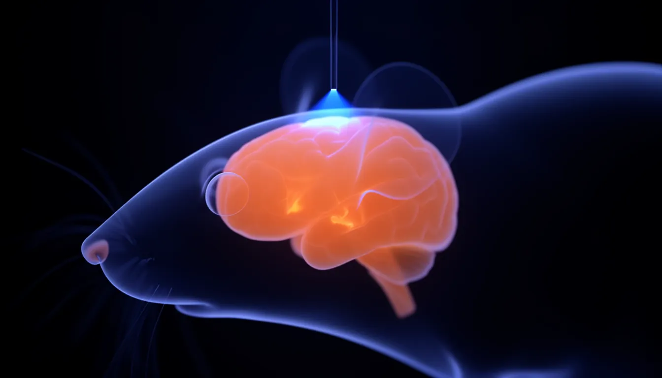
Researchers use viral vectors or genetically modified animals so that only a chosen type of neuron makes the protein. Light delivered through an optical fiber then switches just those cells on or off at precisely chosen moments.
What was new
Electrical stimulation excites every cell near the electrode regardless of type. Drugs are hard to target to one cell type and act slowly. Optogenetics offers both cell-type specificity and millisecond timing.
That made it possible to test cause and effect directly, asking whether activity in a given set of neurons produces a given behavior.
History
In 2005 Edward Boyden, Feng Zhang, Karl Deisseroth and colleagues expressed channelrhodopsin-2 in mammalian neurons and showed that light could trigger action potentials on a millisecond timescale. Inhibitory tools and variants tuned to other wavelengths followed, and Deisseroth reviewed the field in Nature Methods in 2011.
Combining with other methods
Optogenetics becomes more powerful when paired with techniques that read out brain activity. Optogenetic fMRI (ofMRI), which combines optogenetic stimulation with fMRI, maps which circuits across the whole brain respond when specific cells are driven. The method was published in Nature in 2010 by Jin Hyung Lee and colleagues.
Limits
Using optogenetics in people requires both gene delivery and light delivery, so it is mainly used today as a tool for uncovering how neural circuits work in animal research.
Related entries
- Optogenetic fMRI (ofMRI) — Optogenetic fMRI (ofMRI) drives specific neurons with light while measuring whole-brain responses with fMRI, mapping the circuits those cells recruit.
- Neural circuit — A neural circuit is a group of neurons connected by synapses that work together to carry out a specific function such as sensory processing, movement or memory.
- Functional magnetic resonance imaging (fMRI) — fMRI uses MRI to track blood-oxygen changes (the BOLD signal) that follow neural activity, showing noninvasively which brain regions are active.
References
- Millisecond-timescale, genetically targeted optical control of neural activity. Boyden ES, Zhang F, Bamberg E, Nagel G, Deisseroth K. Nature Neuroscience 8, 1263–1268 (2005)
- Optogenetics. Deisseroth K. Nature Methods 8, 26–29 (2011)
- Global and local fMRI signals driven by neurons defined optogenetically by type and wiring. Lee JH et al. Nature 465, 788–792 (2010)