Optogenetic fMRI (ofMRI)
Also known as: ofMRI, opto-fMRI
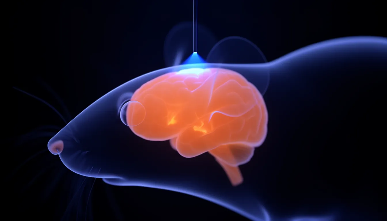
In one sentenceOptogenetic fMRI (ofMRI) drives specific neurons with light while measuring whole-brain responses with fMRI, mapping the circuits those cells recruit.
Overview
ofMRI asks how the whole brain responds when you know exactly which cells you switched on. Optogenetics supplies the cause; fMRI measures the effect.
The 2010 Nature paper
The method was established in the 2010 Nature paper “Global and local fMRI signals driven by neurons defined optogenetically by type and wiring.” Jin Hyung Lee, now a Stanford professor, was first author and Karl Deisseroth was senior author.
The team expressed channelrhodopsin-2 in excitatory (CaMKIIα-expressing) neurons of rodent motor cortex and stimulated them with 473 nm blue light. Robust BOLD signals appeared in the stimulated cortex and in distant connected regions, while control animals without the gene showed no such signals.
Two points followed:
- Activity in defined excitatory neurons is enough to produce a BOLD signal.
- It is possible to see, in a living animal, which brain-wide circuits a local stimulus recruits.
Why it matters
fMRI is the workhorse of human brain research, but the cellular source of its signal could only be inferred. Because ofMRI fixes the cause before measuring the effect, it provides a reference point for interpreting fMRI. It is also used to look for disease-related neural circuits in animal models.
Later developments
Lee’s lab at Stanford describes one of its main themes as “From ofMRI to Mechanogenetic Functional Ultrasound.” The work funded by Lee’s 2019 NIH Director’s Pioneer Award develops mechanogenetics, which perturbs cells in the brain mechanically without surgery, and functional ultrasound imaging of whole-brain activity in awake, moving animals.
Related entries
- Optogenetics — Optogenetics delivers light-sensitive proteins to chosen neurons so researchers can switch those cells on or off with light, with millisecond precision.
- Functional magnetic resonance imaging (fMRI) — fMRI uses MRI to track blood-oxygen changes (the BOLD signal) that follow neural activity, showing noninvasively which brain regions are active.
- Neural circuit — A neural circuit is a group of neurons connected by synapses that work together to carry out a specific function such as sensory processing, movement or memory.
- Jin Hyung Lee (neuroscientist) — Jin Hyung Lee is a Stanford associate professor of neurology, neurosurgery and bioengineering, known for optogenetic fMRI (ofMRI) and brain circuit research.
References
- Global and local fMRI signals driven by neurons defined optogenetically by type and wiring. Lee JH, Durand R, Gradinaru V, Zhang F, Goshen I, Kim DS, Fenno LE, Ramakrishnan C, Deisseroth K. Nature 465, 788–792 (2010)
- Stanford University Lee Lab — Research Themes
- $4.75 million awarded to scientists for high-risk, high-reward research. Stanford Medicine News Center, 2019-10-01