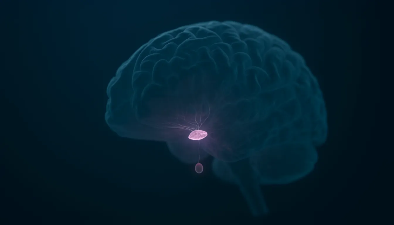
Hypothalamus
Also known as: hypothalamus function, hypothalamus location, what does the hypothalamus do, hypothalamic nuclei, hypothalamus and pituitary gland, suprachiasmatic nucleus, arcuate nucleus, preoptic area
In one sentenceThe hypothalamus is a small region at the base of the brain, below the thalamus, that keeps the body's internal state stable through the autonomic nervous system and hormones.
What does the hypothalamus do?
The hypothalamus is a small structure deep in the brain, about the size of an almond, whose main job is to keep the body in a stable internal state, called homeostasis. It works in two ways: by directly influencing the autonomic nervous system, which runs automatic functions such as heart rate and breathing, and by managing hormones. The Queensland Brain Institute describes it as the major control centre of the autonomic motor system and as the part of the brain that connects the hormonal and nervous systems, and a 2023 review calls it the highest regulator of the autonomic nervous system.
Among the things it helps regulate are body temperature, hunger and thirst, blood pressure, sleep and daily rhythms, mood and sex drive. The sections below set out where it is, how it is organized, and the research behind each of these roles.
Location and name
The hypothalamus is part of the diencephalon. It lies below the thalamus and above the midbrain; as its name suggests, it sits right underneath the thalamus. The pituitary gland sits just below it. Anatomical landmarks that mark out its regions include the lamina terminalis, the pituitary gland, the mammillary bodies and the superior hypothalamic sulcus.
Claudius Galen in the second century and Andreas Vesalius in the 16th century described the brain region corresponding to part of the hypothalamus, but Wilhelm His coined the name “hypothalamus” in 1893.
Nuclei and connections
The hypothalamus is not one uniform piece of tissue. It is a collection of nuclei, clusters of neurons, on both sides of the brain, arranged in three zones around the third ventricle and the mammillary bodies. Nuclei in the periventricular zone generally regulate the endocrine system, while those in the medial and lateral zones regulate autonomic and somatic behavior.
Named nuclei include the paraventricular, ventromedial, dorsomedial, preoptic, supraoptic, suprachiasmatic and arcuate nuclei, along with the lateral hypothalamic area. They are connected to one another and to many surrounding brain regions. Among the main pathways, the hypothalamus connects:
- to the brainstem through the dorsal longitudinal fasciculus,
- to the cerebral cortex through the medial forebrain bundle,
- to the hippocampus through the fornix,
- to the amygdala through the stria terminalis,
- to the thalamus through the mammillothalamic tract,
- to the pituitary through the median eminence, and
- to the retina through the retinohypothalamic tract.
Link to the pituitary gland
The pituitary gland has two lobes, anterior and posterior, and the hypothalamus reaches them in different ways. It communicates with the anterior lobe through a network of blood vessels and with the posterior lobe through the pituitary stalk.
The hypothalamus makes two hormones itself, oxytocin and vasopressin, and stores them in the posterior pituitary, which releases them into the bloodstream when they are needed. Oxytocin stimulates the uterine muscle to contract during birth and is involved in the release of breast milk. Vasopressin, also called antidiuretic hormone, regulates the body’s water and urine volume and blood pressure.
The control of the anterior lobe was worked out later. It had long been known that the nervous system could in some way influence the endocrine glands, probably with the hypothalamus acting as the go-between, and as early as 1930 small blood vessels had been found connecting the hypothalamus with the pituitary. Toward the end of the 1950s Roger Guillemin and Andrew Schally, working in separate laboratories, extracted compounds from the hypothalamus of sheep and pigs that made pituitary tissue release its hormones. They called these substances releasing factors or releasing hormones.
Their chemical nature was not established until 1969. Guillemin worked with 5 million hypothalamic fragments from sheep and Schally with the same amount of material from pigs, and after years of work they had 1 mg of a pure substance that released thyroid-stimulating hormone from the pituitary. This was TRF, the TSH-releasing factor, an extremely small peptide made of three amino acids. Within two years the releasing hormone for luteinizing hormone, LH-RH, had also been isolated, sequenced and synthesized. Further peptides followed, including somatostatin, the first known inhibitor of pituitary function, which reduces the pituitary’s production of growth hormone. Guillemin and Schally shared half of the 1977 Nobel Prize in Physiology or Medicine for their discoveries concerning “the peptide hormone production of the brain”.
Several of these signals come from neuroendocrine neurons in the arcuate nucleus. They release gonadotropin-releasing hormone, which stimulates the anterior pituitary to make FSH and LH, and growth hormone-releasing hormone, which stimulates the secretion of growth hormone. Work in the 1960s and 1970s reported that dopamine released by arcuate neurons regulates the pituitary’s secretion of prolactin.
Hunger and body weight
The arcuate nucleus sits in the lower middle part of the hypothalamus, next to the third ventricle and the median eminence. A 2023 review by Juhyun Song and Seok-Yong Choi describes it as the center where nutrition, metabolism and reproduction are kept in balance.
Two intermingled groups of arcuate neurons pull in opposite directions. Neurons that make agouti-related peptide (AgRP) and neuropeptide Y promote eating. They are activated by ghrelin and switched off by leptin, insulin and glucose in the blood. Leptin is a hormone produced mainly by fat cells that reduces appetite. Neurons that make pro-opiomelanocortin (POMC) respond to leptin by producing α-MSH, which acts on receptors called MC4R in the paraventricular nucleus. AgRP binds the same family of receptors and suppresses the appetite-reducing effect of α-MSH.
In 2011 Yexica Aponte, Deniz Atasoy and Scott Sternson tested how much these neurons can do on their own. They used optogenetics, making AgRP or POMC neurons in mice responsive to light with the protein channelrhodopsin-2. Switching on only 800 AgRP neurons made the mice eat voraciously within minutes, and the response grew with the number of neurons stimulated and the frequency and duration of the light. Stimulating POMC neurons instead reduced food intake and body weight. The AgRP-driven feeding appeared without any training and was selective for eating over drinking, which the authors took to mean that these neurons have a dedicated role in coordinating feeding.
Body temperature
The body’s core temperature is held within a defended range by a combination of feedback, triggered when core temperature drifts, and feed-forward signals such as skin thermoreceptors detecting a change in air temperature before the core is affected. That defended value, sometimes called the set point, is not fixed but shifts with internal and external conditions.
According to a 2018 review by Chan Lek Tan and Zachary Knight, the preoptic area of the hypothalamus is thought to be the key site in the brain that integrates this information. The evidence includes the following:
- animals with a damaged or silenced preoptic area cannot defend their core temperature in either a hot or a cold environment;
- local warming of the preoptic area lowers body temperature and produces the same heat-defense responses as a hot environment, while cooling the most temperature-sensitive spot in it does the opposite, promoting constriction of blood vessels, heat production in brown fat, shivering and heat-seeking behavior;
- injecting the fever-causing molecule prostaglandin E2 into the preoptic area causes fever;
- the area contains neurons that are activated by warming of the skin, spinal cord or brain.
Fever itself works through this region. Immune cells that detect bacteria or viruses produce cytokines and prostaglandins that act on the preoptic area, and prostaglandin E2 acts through EP3 receptors in the median preoptic nucleus to change body temperature.
Sleep and daily rhythms
The suprachiasmatic nucleus acts as a pacemaker for the body’s daily, or circadian, rhythms. In 1972 Friedrich Stephan and Irving Zucker found that destroying the suprachiasmatic nuclei on both sides in rats permanently eliminated the animals’ 24-hour rhythms of drinking and moving about, while destroying the medial preoptic area had no effect on the drinking rhythm. They suggested that neurons in the suprachiasmatic region may coordinate both the generation of these rhythms and their alignment with the cycle of light and dark.
A 1990 transplant experiment showed that cells of the suprachiasmatic region set the rhythm’s basic period. Martin Ralph, Michael Menaker and colleagues used a mutant strain of hamster with a short circadian period. Small grafts from the suprachiasmatic region restored rhythms to animals whose own nucleus had been removed, and the restored rhythm always ran at the period of the donor’s genotype, not the host’s.
The front of the hypothalamus and the neighboring basal forebrain take part in generating sleep. In 1996 Sherin, Saper and colleagues identified a group of neurons in the ventrolateral preoptic area that become active specifically during sleep. These neurons send connections to the tuberomammillary nucleus, a group of cells in the back of the hypothalamus thought to take part in arousal, and the authors proposed that this pathway may play a key role in determining whether an animal is asleep or awake.
In 1998 two research groups reported the same pair of neuropeptides, made by neurons in and around the lateral hypothalamus, under different names. De Lecea, Sutcliffe and colleagues named them hypocretins and found that the neurons’ fibers reach the brainstem and thalamus and that one of the peptides excited hypothalamic neurons. Takeshi Sakurai, Masashi Yanagisawa and colleagues named them orexins and found that they increased food intake when given to rats and that their gene was more active during fasting. These cells are also studied in research on narcolepsy, a sleep disorder. Narcolepsy in mice and dogs can be caused by mutations in the gene for the hypocretin precursor or its receptors, but most human narcolepsy does not run in families and has not been linked to mutations in this system. In 2000 Thomas Thannickal, Jerome Siegel and colleagues found that brains of people who had narcolepsy had 85–95% fewer hypocretin neurons, while a neighboring type of neuron was not reduced, a pattern the authors considered consistent with a degenerative process.
Defense and aggression
Walter Rudolf Hess received half of the 1949 Nobel Prize in Physiology or Medicine “for his discovery of the functional organization of the interbrain as a coordinator of the activities of the internal organs”. The interbrain, or diencephalon, includes the hypothalamus. Hess placed fine wires, insulated except at the tip, into very small areas of the brains of anesthetized cats, and once the animals had fully recovered he passed a weak current through them. Depending on where the tip lay, a cat would curl up and fall into a sleep from which it could be woken as easily as from natural sleep, or would react as if threatened by a dog: its hair stood on end, it spat and, if the stimulus continued, it attacked. Other sites changed circulation and breathing. The responses combined autonomic reactions with fitting movements of the body, such as postures and attempts to escape or attack.
Later work narrowed down where aggression is controlled. Electrical stimulation of certain hypothalamic regions in cats and rodents was known to trigger attack, but the exact cells involved were unclear. In 2011 Dayu Lin, David Anderson and colleagues showed that optogenetic stimulation, though not electrical stimulation, of neurons in the ventrolateral part of the ventromedial hypothalamus made male mice attack females, males and even inanimate objects. Silencing these neurons reversibly reduced fighting between males. Recordings showed overlapping but distinct groups of neurons active during fighting and during mating, and neurons active during attack were inhibited during mating.
Related entries
- Amygdala — The amygdala is an almond-shaped cluster of nuclei deep in each temporal lobe, next to the hippocampus. It links sights and sounds with emotional value, above all threat.
- Hippocampus — The hippocampus is a structure deep in the temporal lobe that is central to turning new events and facts into long-term memories and to remembering places.
- Dopamine — Dopamine is a neurotransmitter for movement, motivation and reward learning. Often called the pleasure chemical, it mainly signals prediction errors.
- Optogenetics — Optogenetics delivers light-sensitive proteins to chosen neurons so researchers can switch those cells on or off with light, with millisecond precision.
References
- Forebrain. Queensland Brain Institute, The University of Queensland
- The limbic system. Queensland Brain Institute, The University of Queensland
- Neuroanatomy, Hypothalamus. Bear MH, Reddy V, Bollu PC. StatPearls, NCBI Bookshelf
- Hypothalamus. Cleveland Clinic, medically reviewed 16 March 2022
- Arcuate nucleus of the hypothalamus: anatomy, physiology, and diseases. Song J, Choi SY. Experimental Neurobiology 32, 371–386 (2023)
- The Nobel Prize in Physiology or Medicine 1977 — Press release. Karolinska Institutet, October 1977
- The Nobel Prize in Physiology or Medicine 1949 — Presentation speech. Olivecrona H, Royal Caroline Institute
- AGRP neurons are sufficient to orchestrate feeding behavior rapidly and without training. Aponte Y, Atasoy D, Sternson SM. Nature Neuroscience 14, 351–355 (2011)
- Regulation of body temperature by the nervous system. Tan CL, Knight ZA. Neuron 98, 31–48 (2018)
- Circadian rhythms in drinking behavior and locomotor activity of rats are eliminated by hypothalamic lesions. Stephan FK, Zucker I. PNAS 69, 1583–1586 (1972)
- Transplanted suprachiasmatic nucleus determines circadian period. Ralph MR, Foster RG, Davis FC, Menaker M. Science 247, 975–978 (1990)
- Activation of ventrolateral preoptic neurons during sleep. Sherin JE, Shiromani PJ, McCarley RW, Saper CB. Science 271, 216–219 (1996)
- The hypocretins: hypothalamus-specific peptides with neuroexcitatory activity. de Lecea L et al. PNAS 95, 322–327 (1998)
- Orexins and orexin receptors: a family of hypothalamic neuropeptides and G protein-coupled receptors that regulate feeding behavior. Sakurai T et al. Cell 92, 573–585 (1998)
- Reduced number of hypocretin neurons in human narcolepsy. Thannickal TC, Moore RY, Nienhuis R, Ramanathan L, Gulyani S, Aldrich M, Cornford M, Siegel JM. Neuron 27, 469–474 (2000)
- Functional identification of an aggression locus in the mouse hypothalamus. Lin D, Boyle MP, Dollar P, Lee H, Lein ES, Perona P, Anderson DJ. Nature 470, 221–226 (2011)