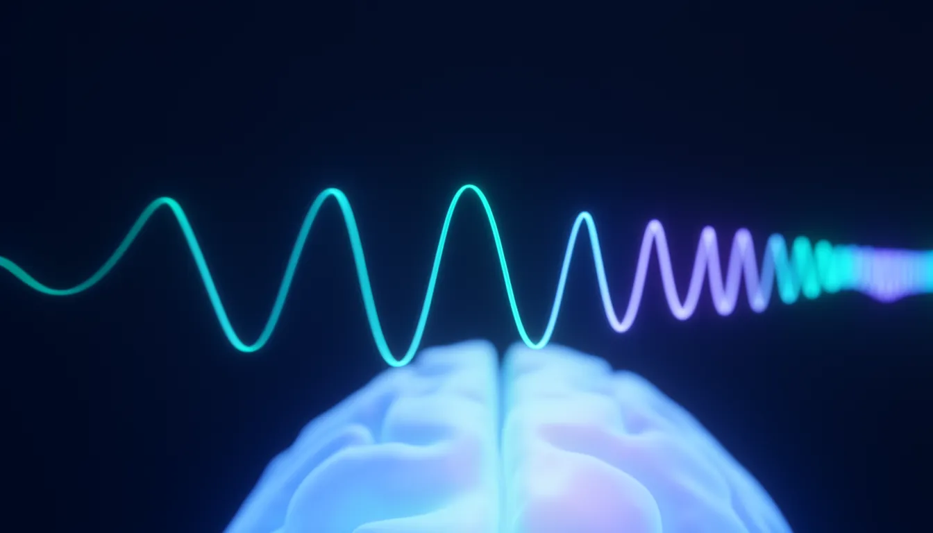
Electroencephalography (EEG)
Also known as: EEG, brain waves, electroencephalogram
In one sentenceEEG records the brain's electrical activity through scalp electrodes with millisecond precision. It is used in sleep research and in epilepsy evaluation.
What it measures
A single neuron’s electrical signal is far too weak to reach the scalp. EEG measures the summed electric field produced when synaptic currents flow at the same time in vast numbers of cortical neurons. EEG therefore shows how well many cells are keeping time together, not what any one cell is doing.
History
German psychiatrist Hans Berger recorded the human EEG in 1924 and published the results in 1929. The 8–13 Hz rhythm that appears clearly when a person rests with eyes closed is still called the alpha rhythm, or the Berger rhythm.
Frequency bands
Boundaries vary slightly between studies, but bands are roughly divided as follows.
| Band | Frequency | Often seen during |
|---|---|---|
| Delta | below 4 Hz | deep sleep |
| Theta | 4–8 Hz | drowsiness, some memory tasks |
| Alpha | 8–13 Hz | relaxed rest with eyes closed |
| Beta | 13–30 Hz | alert concentration |
| Gamma | above 30 Hz | sensory processing, cognitive tasks |
Strengths and limits
EEG has excellent time resolution, catching changes within milliseconds, and the equipment is relatively small and inexpensive. Because the signal spreads as it passes through the skull and scalp, its spatial resolution, knowing exactly where a signal came from, is low. It complements fMRI, which has high spatial resolution but low time resolution.
Uses
EEG is used to score sleep stages in sleep research, in neurological tests such as epilepsy evaluation, and in brain–computer interface research. Reading and interpreting EEG recordings takes specialized training.
Related entries
- Functional magnetic resonance imaging (fMRI) — fMRI uses MRI to track blood-oxygen changes (the BOLD signal) that follow neural activity, showing noninvasively which brain regions are active.
- Neural circuit — A neural circuit is a group of neurons connected by synapses that work together to carry out a specific function such as sensory processing, movement or memory.
- Synapse — A synapse is the junction where one neuron passes a signal to the next. Most are chemical synapses that relay the signal by releasing neurotransmitters.
References
- Über das Elektrenkephalogramm des Menschen. Berger H. Archiv für Psychiatrie und Nervenkrankheiten 87, 527–570 (1929)
- The origin of extracellular fields and currents — EEG, ECoG, LFP and spikes. Buzsáki G, Anastassiou CA, Koch C. Nature Reviews Neuroscience 13, 407–420 (2012)
- Principles of Neural Science, 6th ed.. Kandel ER, Koester JD, Mack SH, Siegelbaum SA (eds). McGraw Hill (2021)