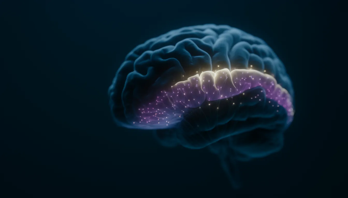
Cortical homunculus
Also known as: homunculus, motor homunculus, sensory homunculus, somatosensory homunculus, Penfield homunculus, homunculus brain
In one sentenceThe cortical homunculus is a distorted human figure that pictures how the body is mapped along the brain's motor and touch strips. Hands, mouth and tongue look huge.
What is the cortical homunculus?
The cortical homunculus, whose name means “little man”, is a drawing of a human body laid out along a strip of the brain’s surface, with each body part scaled by how much cortex is devoted to it rather than by its real size. That is why it has long fingers, a large mouth and an oversized tongue. There are two closely linked versions. The motor homunculus pictures the strip that controls movement, and the sensory (somatosensory) homunculus pictures the strip that receives touch. The neurosurgeon Wilder Penfield and his colleague Edwin Boldrey introduced the figure in 1937. It is a teaching aid, not a precise anatomical map, and later research has refined it in several ways described below.
Where the two maps lie
The two maps sit side by side, one on each side of the central sulcus, the groove that separates the frontal lobe from the parietal lobe behind it.
- Motor map. It lies on the precentral gyrus, the ridge just in front of the central sulcus. This is the primary motor cortex, the frontal lobe’s main region for voluntary movement.
- Sensory map. It lies on the postcentral gyrus, the ridge just behind the groove, in the parietal lobe, which integrates touch, temperature, pressure and pain.
Along both strips the body is laid out in a rough order. The legs and feet are represented near the top of the brain, the arm, hand and fingers further down the side, and the face, mouth and tongue lowest of all. Each hemisphere’s maps serve mainly the opposite side of the body: in a 2018 stimulation study the hand was mapped in the hemisphere opposite to it.
How Penfield made the map
Penfield and Boldrey’s 55-page paper appeared in the journal Brain on 1 December 1937. It drew on electrical stimulation of the cortex in 126 patients, including people with hard-to-treat epilepsy, whom Penfield operated on under local anaesthesia between 1928 and 1936. Because the patients were awake, they could report the sensations and movements each stimulation produced. Boldrey sketched 170 summary maps of the stimulation points for each body part from Penfield’s notes, photographs and drawings, and the homunculus condensed them into one figure.
According to Marco Catani’s 2017 reanalysis of the original data, the figure was introduced after 42 pages and 27 figures, with only a few lines of text. The original showed the man upside down, with his head detached from his body. In a 1950 publication with Rasmussen, Penfield replaced it with a version of better proportions and corrected the exaggerated tongue, while warning that such drawings “may easily become confusing if too much significance is attributed to the shape and comparative size”.
Why the hands and lips are so large
The usual explanation is that the amount of cortex given to a body part depends on how much nerve supply links it with the cortex, not on how big the part is. Body parts with highly skilled movement or perception receive more of that supply, so the proportions of the homunculus are generally thought to reflect specialization of function.
This shows up in touch. Telling whether one or two nearby points are touching the skin, called two-point discrimination, is easier on some parts of the body than others because they have more sensory receptors, and it is generally easier on the parts that are enlarged in the sensory homunculus. In the 2018 stimulation study by Franck-Emmanuel Roux and colleagues, the current needed to produce a sensation was lower for the hand and tongue than for other body parts.
What the drawing leaves out
Catani’s reanalysis found that Penfield and Boldrey’s own data were messier than the neat figure suggests.
- Overlap. Stimulation sites for different body parts overlapped widely, especially near the central sulcus, and the same body part could be reached from sites far apart. The central sulcus was not a clean border either: many motor responses came from the sensory strip, and an even greater proportion of touch responses came from the motor strip.
- Scaling. The original figure was scaled by the vertical length of each body area along the strip, not by its surface area, and its proportions did not match the paper’s own measurements. The tongue, for example, was clearly exaggerated.
- Variation between people. Data from many patients were pooled without statistical weighting, and the clean, orderly pattern has rarely been replicated in a single patient.
- Whose body. Penfield mapped both men and women, but he reported little about his female patients, and the famous figure is unmistakably male. In 2013 Di Noto and colleagues reviewed how much remains unknown about how the female body is represented in the sensory cortex.
Revising the map
Later studies have tested the homunculus with newer methods.
- A modern touch map. In the 2018 study, Roux and colleagues stimulated the postcentral gyrus of 50 patients during surgery and recorded the positions in a standard brain coordinate space. Of 647 sites stimulated in one part of the strip (Brodmann area 1), 258 (40%) produced a sensation. The order of body parts from the top to the side of the brain varied little between patients, and within the hand the tip of each finger lay further back than its base. Unlike Penfield, they found no sensations from outside the postcentral gyrus, no sense of movement or desire to move, and no responses on both sides of the body from face or tongue stimulation. They proposed a redrawn sensory homunculus.
- Maps of actions. In 2002 Michael Graziano and colleagues stimulated the motor and neighboring premotor cortex of monkeys for half a second at a time, about as long as a natural reach or grasp. The stimulation evoked coordinated postures involving many joints. At one site the mouth opened while the hand formed a grip and moved to the mouth. Sites that moved the arm were arranged as a map of hand positions around the body.
- An interrupted homunculus. In 2023 Evan Gordon, Nico Dosenbach and colleagues scanned seven people for about three to six hours each with precision fMRI and checked the results in large datasets, including the UK Biobank, the ABCD study and the Human Connectome Project. Along the motor strip, separate regions for the foot, hand and mouth alternated with three “inter-effector” regions that had thinner cortex and were strongly functionally connected to each other and to the cingulo-opercular network, a network linked to action, physiological control and arousal. These regions did not prefer any single movement. They became active when people planned coordinated movements of the hands and feet or moved the abdomen or eyebrows. The authors called this system the somato-cognitive action network (SCAN) and proposed that the motor strip combines two systems: one for isolated fine control of the foot, hand and mouth, and one for whole-body action planning.
Maps that change with experience
The homunculus is not fixed, a form of neuroplasticity.
- After losing a finger. In 1984 Michael Merzenich and colleagues mapped the hand area of the touch cortex in adult owl monkeys 2 to 8 months after one or two fingers had been surgically removed. The areas for the neighboring fingers and palm had expanded to take over most or all of the cortex that had represented the missing fingers. The major changes were confined to a zone about 0.5–0.7 mm on either side of the old borders.
- In musicians. In 1995 Thomas Elbert and colleagues used magnetic source imaging to compare string players with control participants. The cortical representation of the fingers of the left hand, which finger the strings, was larger in the musicians, while the right hand showed no such difference. The effect was smallest for the left thumb, and its size was related to the age at which the musicians had begun to play.
Related entries
- Frontal lobe — The frontal lobe is the front part of each cerebral hemisphere. It controls voluntary movement, produces speech and, through the prefrontal cortex, plans and regulates behavior.
- Neuroplasticity — Neuroplasticity is the brain's capacity to change synapses and connections with experience, the cellular basis of learning and memory.
- Functional magnetic resonance imaging (fMRI) — fMRI uses MRI to track blood-oxygen changes (the BOLD signal) that follow neural activity, showing noninvasively which brain regions are active.
References
- Somatic motor and sensory representation in the cerebral cortex of man as studied by electrical stimulation. Penfield W, Boldrey E. Brain 60, 389–443 (1937)
- A little man of some importance. Catani M. Brain 140, 3055–3061 (2017)
- Lobes of the brain. Queensland Brain Institute, The University of Queensland
- Functional architecture of the somatosensory homunculus detected by electrostimulation. Roux FE, Djidjeli I, Durand JB. The Journal of Physiology 596, 941–956 (2018)
- Complex movements evoked by microstimulation of precentral cortex. Graziano MSA, Taylor CSR, Moore T. Neuron 34, 841–851 (2002)
- A somato-cognitive action network alternates with effector regions in motor cortex. Gordon EM et al. Nature 617, 351–359 (2023)
- The hermunculus: what is known about the representation of the female body in the brain?. Di Noto PM, Newman L, Wall S, Einstein G. Cerebral Cortex 23, 1005–1013 (2013)
- Somatosensory cortical map changes following digit amputation in adult monkeys. Merzenich MM, Nelson RJ, Stryker MP, Cynader MS, Schoppmann A, Zook JM. Journal of Comparative Neurology 224, 591–605 (1984)
- Increased cortical representation of the fingers of the left hand in string players. Elbert T, Pantev C, Wienbruch C, Rockstroh B, Taub E. Science 270, 305–307 (1995)